NIH grant supports study of the spread of Huntington’s disease in the brain
NIH grant supports study of the spread of Huntington’s disease in the brain
September 13, 2024
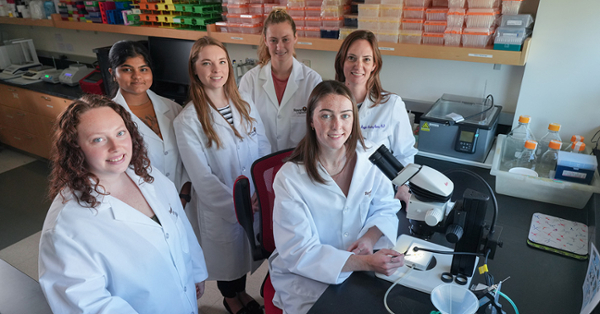
From left, lab members Lilly Boltz, third-year doctoral student; Tanisha Sharma, junior; Dawn Williams, third-year doctoral student; Amy Appollina, senior; Katie Wooster (seated), senior; and Maggie Panning Pearce, Ph.D., are studying how Huntington's disease progresses in the brain.
Just like your house, the brain needs regular cleaning. Cells called glia clear away debris to keep this organ in good working order. But in neurodegenerative disorders such as Huntington’s disease, glia not only fail to do that job, they may also spread the mess.
Through studies conducted primarily with fruit flies (Drosophila melanogaster), Maggie Panning Pearce, Ph.D., an associate professor in the Department of Biological and Biomedical Sciences in the College of Science & Mathematics, determined that this clean-up crew appears to help propagate toxic Huntington’s disease proteins within the brain. Now, with funding from the National Institutes for Health (NIH), she’s working to better understand glial cells’ role in this and other neurodegenerative disorders.
The grant, which she received in 2022 while at another institution, has provided $332,366 for one year of research at Rowan and will likely fund additional years of work on this project.
In Huntington’s disease, a genetic error causes the protein huntingtin to form clumps in the brain, which lead to many neurological symptoms, such as behavioral and cognitive changes, involuntary movement, and difficulty speaking and swallowing. People who inherit the altered gene typically develop these symptoms in middle age. The disease worsens over time, leaving patients unable to care for themselves; doctors currently have no treatment to stop its progression.
Mutations in the gene cause some huntingtin proteins to become misshapen, sparking what researchers think is a cascade reaction. A single malformed molecule can kick it off by causing its normal counterparts to change shape. These aberrant proteins then clump together. As a person ages, the brain becomes less able to rid itself of the malformed protein. So, the aggregates accumulate and damage neurons, brain cells that transmit and receive messages.
Pearce’s research implicates glia, another kind of brain cell. As a postdoc, she demonstrated that glia remove harmful huntingtin aggregates, but that, over time, they lose this ability and instead contribute to spreading them.
“I want to determine, how this is happening?” Pearce says. “If we can figure out how glia control not only the load of aggregates in the brain, but also their ability to spread, maybe we can identify targets for new therapies that could slow disease progression.”
Studies conducted in her lab with this support have used fruit flies to examine how the presence of aggregates may contribute to the glial cells’ switch from helpful to harmful. Her group is also working in flies to identify genes involved in the process by which glia engulf and break down toxic material like misfolded huntingtin. Genes like these could one day become targets for therapies that interfere with the spread of the aggregates.
In another ongoing project, this time using human and mouse cells, the Pearce lab is investigating whether aggregates move from neurons into a type of glial cell called astrocytes.
These experiments have implications beyond Huntington’s. Researchers have documented a similar dynamic, in which malformed proteins seed the formation of toxic aggregates, in other neurodegenerative diseases, including Alzheimer’s, Parkinson’s, frontotemporal dementia and amyotrophic lateral sclerosis.
Students power this research, Pearce says. At any given time, she has four to six undergrads in her lab, working alongside multiple graduate students.
“Flies are very good for teaching students how to perform experiments in genetics, cell and molecular biology, and for discovering the underpinnings of human disease,” she says.